Image Guided Radiation Therapy
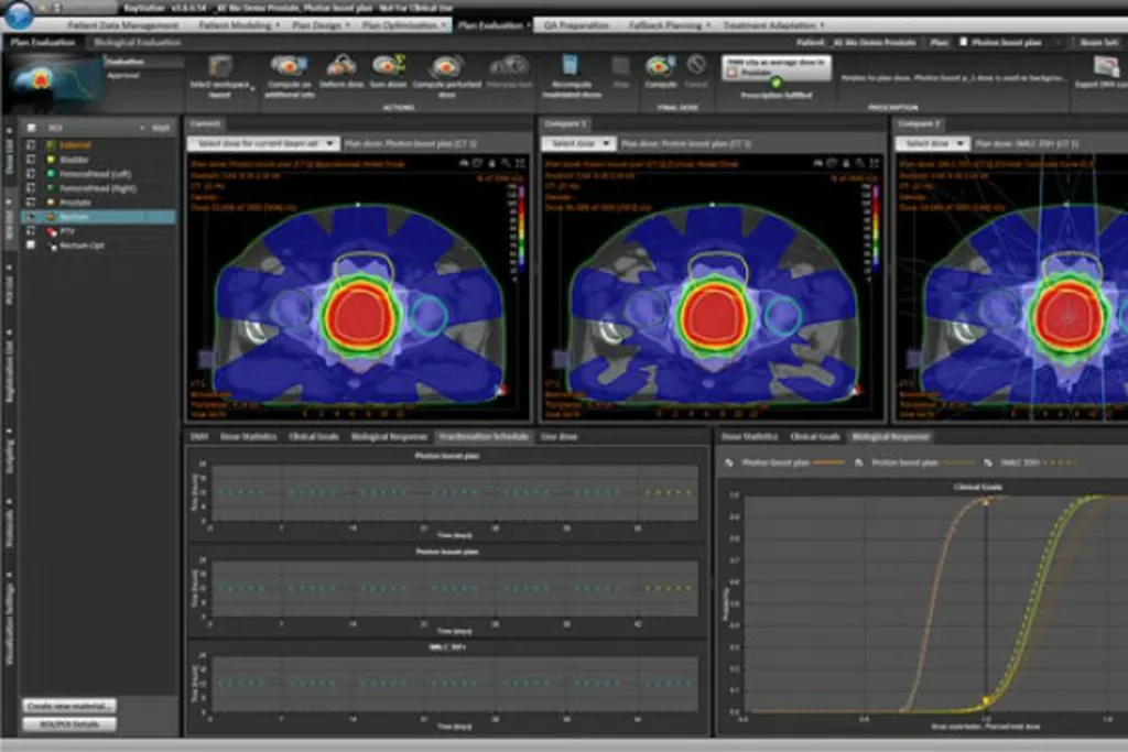
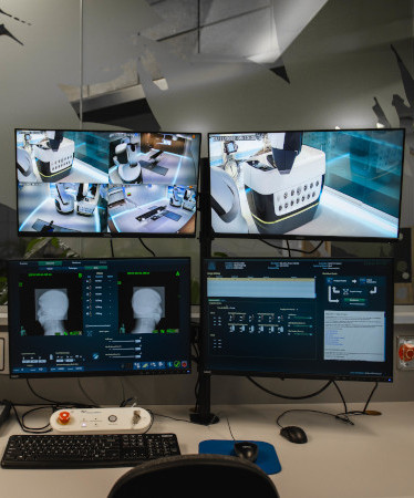
Image guided radiation therapy (IGRT) allows your radiation therapist to see a tumour and how it changes every day. IGRT can help the treatment team target the tumour more accurately and reduce the amount of radiation your healthy tissue receives. It can also cut down on potential side effects. IRGT has made it possible to treat cancers that were hard to access in the past. You may have X-rays and scans before and during your treatment. With these, and a range of 2D, 3D and 4D imaging methods, your treatment team will find, monitor and treat the tumour.
In more detail
Image guided radiation therapy (IGRT) uses a variety of 2D, 3D and 4D imaging techniques during the course of therapy to allow radiographers to see exactly where a tumour is and how it is behaving. This improves the targeting of the radiation therapy, increases the probability of tumour control and spares the normal tissues from undue exposure.
Some tumours may be in a different place after each treatment. IRGT is used to treat tumours in areas of the body where cancers are particularly prone to movement, such as the lungs, liver or prostate gland. To make sure the cancer is inside the radiation therapy field, scans are taken before each treatment. It’s called 4D radiation therapy because they use three dimensional planning, with a fourth dimension of time.
4D is also an excellent technique for targeting tumours close to critical organs. The imaging technologies include XVI 2D and 3D kV which uses planar and volume imaging to allow soft tissue detail to be seen anywhere in the body immediately before treatment.
Elekta Symmetry™ is a unique 4D kV technology that gives volume imaging of moving tumours immediately before treatment. This is matched to the 4D-CT image from the planning phase, and the patient position is corrected to match the plan. This allows moving cancers to be treated aggressively without compromising the safety of surrounding healthy tissue.
Understanding Your Treatment
The Patient Pathway
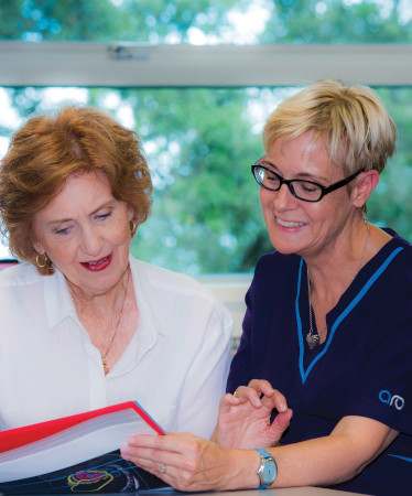
We understand you may be anxious about having radiation therapy. Read about the patient pathway and what to expect before, during and after radiation therapy treatment. AT ARO we work closely with your radiation oncologist to develop an individualised treatment plan for each patient.
Explore the patient pathway